TCD for Sickle Cell Patients: Life-Saving Early Detection
Sickle cell anemia affects thousands of children each year and comes with life-threatening complications, one of the most serious being an increased risk of stroke. Studies show that children with sickle cell disease are up to 300 times more likely to experience a stroke compared to children without the condition. This makes early detection and routine monitoring essential to preventing devastating outcomes.
One of the most powerful tools in stroke prevention for pediatric sickle cell patients is the Transcranial Doppler (TCD) ultrasound. With advanced systems like the Dolphin TCD from Image Monitoring USA, hospitals and pediatric clinics can take proactive steps to identify risk early, intervene promptly, and ultimately save lives.
Why Stroke Risk is Higher in Sickle Cell Patients
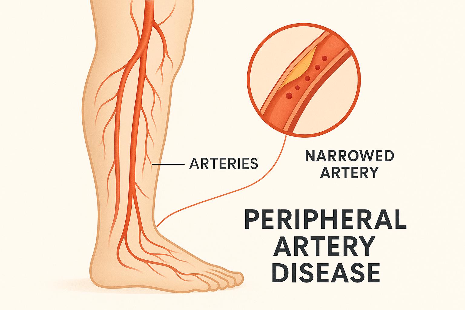
Sickle cell anemia is characterized by abnormally shaped red blood cells that can clump together and block blood vessels. When these blockages occur in the brain’s arteries, they can lead to restricted blood flow or clot formation—both of which dramatically raise the risk of stroke.
Because strokes can occur suddenly and without warning, the medical community emphasizes routine screening as a key preventive measure. Identifying children at highest risk allows providers to begin interventions such as transfusion therapy or other preventive treatments before a stroke occurs.
How TCD Works for Stroke Prevention
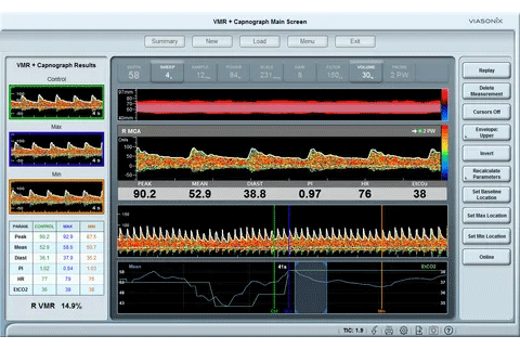
Transcranial Doppler ultrasound is a non-invasive test that measures the velocity of blood flow in major cerebral arteries. In sickle cell patients, elevated flow velocities are a red flag, indicating narrowed vessels and increased stroke risk.
Routine TCD screening in pediatric patients provides critical insights:
- Early identification of high stroke risk through velocity measurements
- Ongoing monitoring to track changes over time
- Immediate, radiation-free results, making it safe for repeated use
- Better outcomes by guiding timely interventions
The non-invasive and radiation-free nature of TCD makes it ideal for children, ensuring their long-term monitoring can be performed safely and frequently.
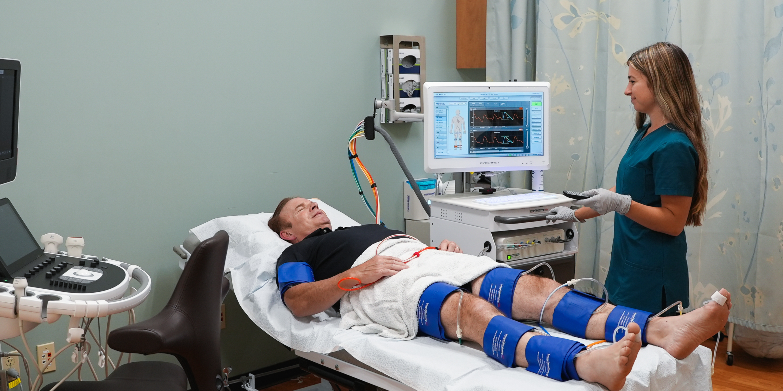
Dolphin TCD: A Vital Tool in Pediatric Care
The Dolphin TCD system from Image Monitoring USA takes stroke prevention in sickle cell patients to the next level. Its advanced features provide:
- Accurate cerebral velocity measurements to detect risk markers early
- Real-time monitoring and reporting for faster clinical decision-making
- Child-friendly, non-invasive testing with no discomfort or radiation exposure
- Ease of use for pediatric teams, ensuring consistent and reliable results
By integrating Dolphin TCD into pediatric care, hospitals can create a proactive strategy that helps children live longer, healthier lives.
Why Every Pediatric Program Should Adopt Routine TCD
National health organizations, including the American Society of Hematology, recommend routine TCD screening for children with sickle cell disease starting at age two. Despite this, not all hospitals have adopted TCD as a standard part of care—leaving vulnerable children at risk.
With its proven ability to detect elevated velocities before a stroke occurs, Dolphin TCD should be a standard diagnostic tool in every pediatric sickle cell program. By identifying risk markers early, physicians can provide life-saving interventions that dramatically reduce the chances of stroke and long-term disability.
Saving Lives Through Early Detection
The reality is simple: stroke prevention in sickle cell patients starts with early detection. TCD gives clinicians the information they need to act before tragedy strikes. By adopting Dolphin TCD, hospitals and pediatric practices are not only improving their standard of care but also giving children the best possible chance at a healthier future.
For clinicians and diagnostic labs, detecting a PFO accurately and quickly is essential. That’s where the Dolphin Transcranial Doppler (TCD) system from Image Monitoring USA comes in.
PFO Detection: Why It Matters
A Patent Foramen Ovale creates a pathway for blood clots or air bubbles to pass from the right side of the heart directly into the brain, bypassing the lungs’ filtering system. This can lead to serious neurological events if undetected. Identifying a PFO not only answers the “why” behind unexplained strokes or migraines but also informs treatment options such as anticoagulation or closure procedures.
Without the right diagnostic tool, PFOs may go unnoticed or be misdiagnosed, leaving patients vulnerable to recurrent, preventable events.
TCD with Contrast: The Gold Standard
While imaging tests like echocardiography are commonly used, Transcranial Doppler with contrast has emerged as the gold standard for right-to-left shunt detection. By monitoring blood flow velocities in cerebral arteries, TCD detects the passage of microbubbles introduced during a contrast injection. When bubbles appear in the cerebral circulation, it confirms the presence of a shunt such as a PFO.
Compared to imaging alternatives, TCD with contrast offers distinct advantages:
- Higher sensitivity in detecting small shunts
- Immediate bedside results, no waiting for scans or interpretation
- Non-invasive and repeatable, without radiation exposure
- Lower cost compared to echocardiography and MRI
Dolphin TCD: Redefining Reliability and Accuracy
The Dolphin TCD system by Image Monitoring USA takes this gold standard even further. Designed for cardiovascular labs and neuro labs, Dolphin TCD combines ease of use with unmatched accuracy. Key features include:
- Reliable right-to-left shunt detection using contrast-enhanced protocols
- Real-time emboli and microbubble monitoring with advanced signal clarity
- User-friendly interface that reduces operator dependency
- Immediate reporting, enabling clinicians to make decisions on the spot
This combination makes Dolphin TCD not just a diagnostic tool, but a critical part of a lab’s stroke prevention and migraine evaluation workflow.
Why Cardiovascular and Neuro Labs Can’t Afford to Miss Dolphin TCD
Every cardiovascular and neuro lab faces the challenge of delivering faster, more precise diagnoses while improving patient outcomes. By incorporating Dolphin TCD, labs can:
- Expand diagnostic capabilities beyond traditional imaging tests
- Improve patient care by detecting PFOs and shunts with accuracy
- Enhance workflow efficiency with immediate, point-of-care results
- Support clinical decisions in managing patients with recurrent TIAs, cryptogenic strokes, or migraine disorders
With PFO present in one in four patients, the ability to rule in or rule out a shunt should be a standard part of modern cardiovascular and neurological care.
The Future of Shunt Detection and Stroke Prevention
As medicine continues to shift toward preventive care and precision medicine, tools like the Dolphin TCD will become indispensable. By giving clinicians rapid, reliable insights into cerebral blood flow and shunt presence, Dolphin TCD helps uncover hidden causes of stroke and migraine—and empowers providers to take action before lasting damage occurs.