Dolphin IQ | 4D | MAX

Advanced Transcranial Doppler Solutions

Advanced Transcranial Doppler Solutions

Dolphin Transcranial Doppler System Supplier

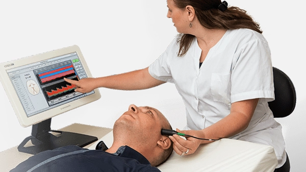
We are the national Dolphin Transcranial Doppler system supplier. The Dolphin TCD system can quickly, non-invasively, and in real-time assess the cerebrovascular system’s function. The Transcranial Doppler “TCD” working principle is based on the Doppler effect and is manufactured as per the laws and regulations governing medical devices. Our customers proudly choose us because we deliver full-fledged clinical applications by offering (TCD) machines with various multi-frequency transducers for use in multiple areas (like Neurology, Radiology, Cardiology, NICU, OR and numerous others), and automatically generated measurement results with trends and graphic reports.

Comprehensive Transcranial Doppler Solutions for Modern Neurovascular Care

Whether you require the all-in-one capabilities of the Dolphin MAX, the advanced diagnostic performance of the Dolphin 4D, or the cost-effective flexibility of the Dolphin IQ every Dolphin system is engineered to support accurate, efficient, and reproducible transcranial Doppler examinations.
IMIUSA
Dolphin IQ

Key Benefits of the
Dolphin 4D TCD System

  • Fully digital Doppler technology delivers best-in-class image and signal quality for more confident diagnoses
  • Review and replay blood flow waveforms with audio at any depth — nothing is missed, even after the exam
  • Unmatched HITS detection with simultaneous multi-depth-gate monitoring catches embolic events other systems may miss
  • High-resolution power M-Mode display gives clearer, more actionable emboli analysis
  • Robotic probe technology enables fast, simple scanning — less operator dependency, more consistent results
  • Comfortable, stable probe fixation with the Dolphin/XF robot saves setup time and reduces technician fatigue
  • Quick-Lock headset design simplifies long-term monitoring setup, freeing staff time
  • Drag & Drop worksheet builder + automatic data feeding lets you examine more patients per day
  • Rated among the most reliable TCD machines on the market — less downtime, more uptime for patient care
  • Slim, lightweight, portable design means the system goes where your patients are, not the other way around
  • Large touch screen is visible from across the room, supporting easier monitoring during procedures
  • Built-in battery (Dolphin/MAX) keeps you working through hours of monitoring without being tied to an outlet
  • Plug-and-play DICOM Worklist & Storage, Structured Report, and HL7 connectivity reduces IT burden
  • Unlimited Review Stations let physicians interpret and report from anywhere — no need to be at the bedside
  • Automatic worksheet transfer to PACS/EMR eliminates manual data entry and transcription errors
  • Dedicated protocols for PFO Bubble Study, Vasospasm Detection, Sickle Cell trending, and Vasomotor Reactivity mean one system covers more of your clinical needs
  • Automatic vasospasm thresholds and day-by-day tracking support earlier, more timely clinical intervention
  • Multi-machine data sharing keeps vasospasm tracking consistent across your institution
  • Step-by-step guided protocols (e.g., Breath Holding Test/BHI) reduce training burden and standardize testing
  • Minimal learning curve with intuitive, large touch-screen software — get new staff up to speed faster
  • Wired/wireless remote and footswitch options keep technicians focused on the patient, not the machine
  • Preconfigured interpretation templates and a user-friendly report builder cut reporting time significantly
  • Custom branding (logo, institute details) delivers a polished, professional patient report
  • Built-in statistics tools, graphing, and Excel integration simplify neuro-vascular research
  • Extensive export options (PDF, Excel, Word, JPEG, VDL, video) support publications, teaching, and presentations
  • Designed as the modern successor to the Sonara TCD system — familiar workflows, dramatically expanded capability
  • Protects your team’s existing investment in TCD expertise while future-proofing your technology
  • Local representatives throughout the USA
  • Multiple RVTs on staff
  • All parts and accessories located in Buffalo NY

IMIUSA Dolphin
4D | MAX

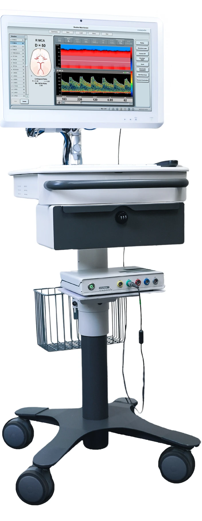
IMIUSA Dolphin IQ

IMIUSA Dolphin IQ… what do you we say about this is should be targetted to explain this model … Lorem ipsum dolor sit amet, consectetur adipiscing elit, sed do eiusmod tempor incididunt ut labore et dolore magna aliqua. Ut enim ad minim veniam.

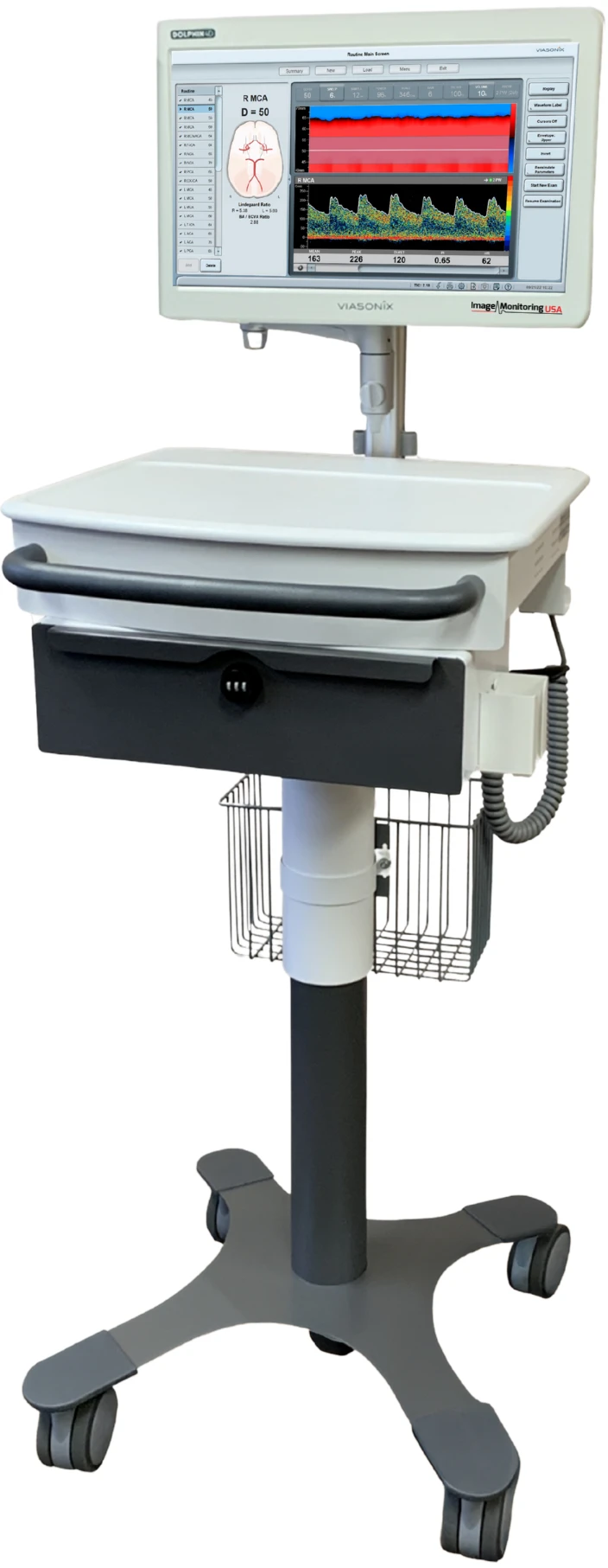
IMIUSA Dolphin 4D | MAX

IMIUSA Dolphin 4D/Dolphin MAX … what do you we say about this is should be targetted to explain this model … Lorem ipsum dolor sit amet, consectetur adipiscing elit, sed do eiusmod tempor incididunt ut labore et dolore magna aliqua. Ut enim ad minim veniam.

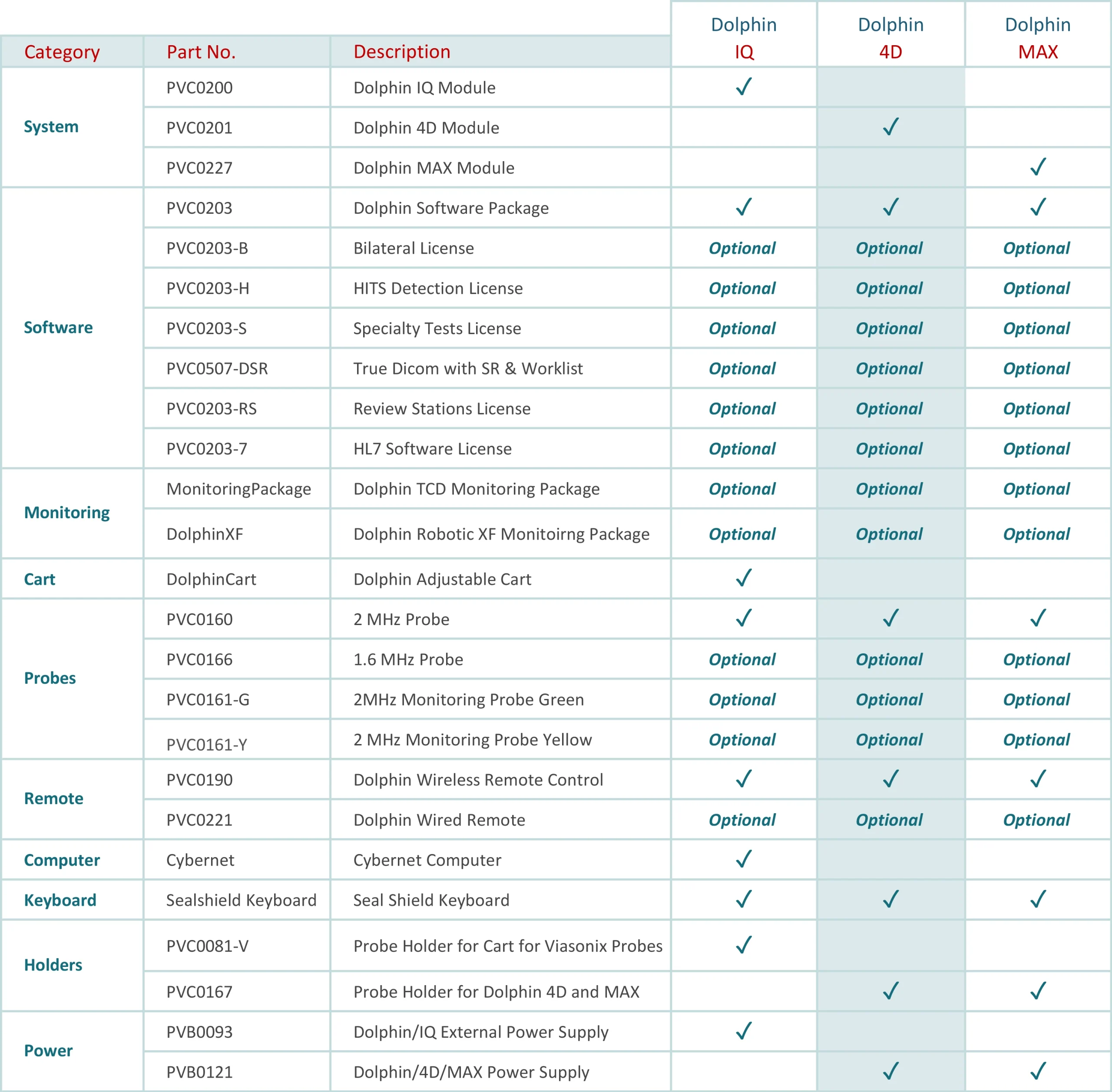
Compare Models

Use our comparison chart to find the Dolphin system best suited to your needs.

Download our comparison chart to find theDolphin system best suited to your needs.

Dowload Dolphin Comparison Chart

Dolphin Enhanced Features

High Sensitivity 2 MHz Pulsed Doppler

The Dolphin’s 2 MHz pulsed-wave probe includes a boost mode for difficult temporal windows, backed by fully digital Doppler processing. Signal quality holds up on the patients who are hardest to insonate, which is exactly where a TCD study is most often abandoned.
Power M-Mode and Multi-Depth Analysis

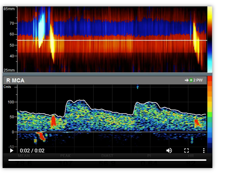
See flow at every depth along the ultrasound beam at once, in both high-resolution color M-mode and the spectral display. Vessel identification is faster, pre- and post-stenotic flow can be compared in the same view, and reversed collateral flow is immediately visible.
Advanced Emboli and HITS Detection

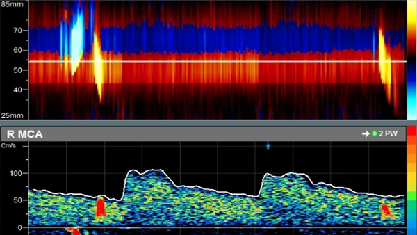
Dedicated HITS analysis screens show each event’s energy, velocity, and duration, along with its travel route through the time and depth domains. Power M-Mode resolves down to 125 microseconds, and multi-range analysis can capture embolic events outside the central measured depth.

Bilateral Monitoring, Hands Free

Adjustable headgear fits quickly and holds bilateral signals through long procedures — cardiac surgery, carotid intervention, or extended bedside monitoring. Automatic and user-placed event markers make post-monitoring review straightforward, and external channel data can be displayed alongside cerebral blood flow.
Dedicated Specialty Protocols

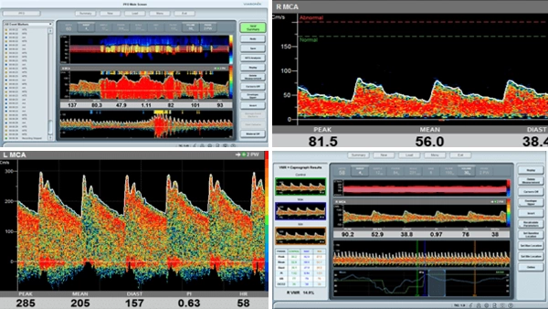
Vasospasm, PFO, sickle cell, vasomotor reactivity, breath holding, and cognitive protocols are built in and ready to run. Each one guides the examiner through the stages of the test and calculates the relevant index automatically, so results are consistent regardless of who performs the study.
Complete Post Editing After the Exam

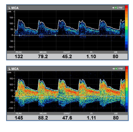
Depth, sample volume, gain, scale, and filter can all be adjusted after the patient has gone home, and any depth can be replayed visually and audibly. Technologists and physicians alike name this the feature they would not give up — few studies ever need to be repeated.
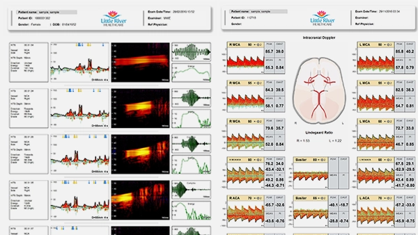
Automatic Statistics, Trends and Reports

Measurements are calculated and tabulated for you, with day-by-day trending that shows exactly when velocities are climbing toward intervention thresholds. Reports are fully configurable, and unlimited review stations let physicians read and post-edit studies remotely through DICOM, DICOM SR, HL7, GDT, and SQL.

A Configuration for Every Department

The Dolphin IQ pairs a 22-inch medical grade touchscreen with a height-adjustable cart and ergo arm for a fixed lab. The 4D is an all-in-one 18-inch standalone that moves easily between units. The MAX adds a six-hour integrated battery for monitoring where power is not guaranteed.

Backed by Our Own Team in Buffalo

Dolphin systems are configured, tested, and shipped from Buffalo, New York, and supported by the same people who sold them. On-site training, application support, and service come from our staff directly — a level of attention a national distributor cannot match.

Dolphin Applications for Transcranial Doppler

Imagine what you would like to have in your ideal TCD system, and discover it in the DOLPHIN!

Cerebral Vasospasm is typically the outcome of Subarachnoid Hemorrhage (SAH) as a result of an aneurysm rupture. Early detection of vasospasm is critical since it allows timely intervention.

TCD is rather specific for this condition, and allows non-invasive, continuous bed-side monitoring.  Vasospasm is characterized by increased velocities due to arterial narrowing. Severe vasospasm is defined in various international guidelines, and typically when mean velocities exceed 200 cm/sec, moderate conditions are with mean velocities between 120-200 cm/sec, and mild vasospasm is for mean velocities near 120 cm/sec. Additionally, velocities in the middle cerebral artery can be compared to internal carotid artery velocities, and the resulting ratio (Lindergaard index) can indicate cerebral vasospasm when it is greater than 3.

Typically, vasospasm is a developing pathology, and requires day by day monitoring of blood flow velocity values.  The IMUSA Dolphin system provides a dedicated vasospasm protocol that allows setting velocity thresholds for immediate identification of the severity of the vasospasm condition.  In addition, an immediate day by day velocity trend analysis is provided in order to help identify the critical time whereby intracranial velocities are dramatically increased, and immediate clinical intervention is required.

The Dolphin also provides an ideal platform for the identification of the critical highest intracranial velocities.  All data is saved both in the time domain and at all depths along the ultrasound beam, which allows the examiner to review the data after freeze to explore the measured blood flow velocities.

Arteriovenous Malformation (AVM) relates to a condition whereby the arterial flow bypasses the capillary circulation and connects directly to the venous drainage, thus not allowing for appropriate tissue perfusion.

This condition is characterized by low resistance to blood flow, resulting in high volume flow rates. AVM is therefore characterized by high mean velocities – although lower then with severe vasospasm – which are coupled with a low pulsatility index (PI).

The IMUSA Dolphin multi-depth options allows to simultaneously view and measure the blood flow spectrum at different depths along the beam and quickly identify deviations from expected waveform patterns.

Peripheral vascular Doppler measurements are normally performed using CW (Continuous Wave) Doppler probes.

Different Probe frequencies are used depending on the target vessels: 4 or 8 MHz are used for larger and deeper vessels such as the Carotid, Femoral or Popliteal arteries, and higher 8 or 10 MHz are used for the smaller and shallower vessels such as the Dorsalis Pedis or Tibial arteries.

The main objective is to qualitatively examine the waveform shapes: a normal peripheral artery (not in the carotids) will show 3 phases during a cardiac cycle, a prominent early systolic forward flow, a late systolic reverse flow, followed by a small component of forward flow again. This signifies a healthy elastic artery. As the vessel becomes less elastic (for example due to diabetes), the third and/or second phase will disappear.

In addition, significantly high velocities may indicate the presence of a stenosis or some other obstruction to the blood flow.

Intracranial stenosis relates to a focal narrowing of the artery, either as a result of an atherosclerotic lesion or as a result of external compression due to a tumor.
TCD will easily identify a l

ocal increase in blood flow velocities, characterized by the fact that proximal or distal to the stenosis the velocities will be much lower that at the site of lesion itself. Peak focal v

elocities above 150 cm/sec can indicate an arterial stenosis. However, if the severity of the stenosis is beyond its’ critical level, velocity signals may be diminished.

The IMUSA Dolphin ideally allows multi-depth scanning along the ultrasound beam, and display in both the m-mode and spectral domains. This allows for simultaneous analysis and evaluation of pre- and post-stenotic flows as well as in the focal region of the stenosis itself.

Vasomotor reactivity (VMR) and autoregulation define the physiological compensatory mechanisms when flow is impaired. Identification of exhausted VMR can be a good indication for high risk for stroke. VMR can be assessed with TCD under two specific conditions: hypercapnia or CO2 reactivity, and with intravenous injections of Acetazolamide or the Diamox test. Note that the Diamox test is not accepted in some countries.

Typically, an increase in peak velocities above 40% during induced increase in CO2 reflects intact VMR, whereas a smaller increase reflects different degrees of impaired VMR. Absence of flow increase reflects a complete exhaustion of the autoregulatory capacity.

The IMUSA Dolphin is ideally set with dedicated Unilateral and Bilateral protocols to determine the autoregulatory capacity of the cerebral circulation of the patient. Doppler waveforms are captured automatically during baseline, and peak (hypercapnia) and minimal (hypocapnia) flows for immediate evaluation, as well as a display of the blood flow fluctuations during entire test process. The respective parameters are automatically shown in a dedicated table with the VMR index readily displayed.

The Breath Holding (BH) test is a specific test for evaluation of Vasomotor Reactivity (VMR) and cerebral autoregulatory capacity.

Identification of exhausted VMR can be a good indication for high risk for stroke. Breath holding induces and increase in CO2, which in turn results in cerebral vasodilation. An increase in peak velocities during breath holding indicates different levels of patent autoregulatory capacity, While the absence of flow increase during the BH test indicates diminished physiological vasodilatory capacity and potentially high risk for stroke.

The IMUSA Dolphin is ideal for BH test with a dedicated breath holding protocol. This protocol leads the examiner with the different stages of the test, and provides automatically the breath holding index (BHI) for clinical determination.

A large timer displayed during the test helps both the patient and the examiner in keeping track of the duration of the procedure. Automatically placed markers identify the peak and minimal velocities measured during the procedure.

Cerebral monitoring can be critical for identification of neurological complications. Relative and/or sudden changes in cerebral blood flow velocities can become a powerful diagnostic tool and effect the immediate clinical intervention.

Bilateral cerebral monitoring is particularly useful during cardiac surgery for identification of embolic events, which may be the source of post-surgery neurological deficiencies.

The IMUSA Dolphin supports both unilateral and bilateral cerebral blood flow monitoring with a dedicated and easy to use monitoring headset. A variety of monitoring tools allow to view the complete duration of the monitored procedure, as well as zoom in or out on specific regions of interest. A variety of automatically inserted event markers, as well as user defined markers, help in post-monitoring analysis.

In addition, the Dolphin supports the display of data obtained from external channels for close correlation with the cerebral blood flow information.

Cerebral emboli can take the form of atherosclerotic plaques, air bubbles or blood clots. An embolus has distinct characteristics which allow to identify it when it travels within the blood stream. The risk of cerebral embolization is in immediate impaired perfusion to the tissue of the brain, which may lead to stroke and sudden death.

TCD can detect the embolic signals in the blood flow velocity waveforms, which are typically characterized with:
• High energy
• Unidirectional
• Traveling within the blood spectrum
• Short in duration
• Typical “chirping” or “bleeping” sound

High intensity transient signals (HITS) are often considered as embolic events. The IMUSA Dolphin has the most advanced HITS detection and analysis platform. Dedicated HITS Analysis screens show the specific embolic characteristics, such as its energy, velocity and duration patterns, as well as display its travel route in the time and depth domains. A special Power M Mode with an Ultra-high time resolution provides the option to zoom in on the m-mode screen at 125 micro-second resolution.

Furthermore, the Dolphin can optionally detect embolic events that are even not within the measured spectrum of the central depth, due to its powerful multi-range and multi-depth analysis capabilities.

Transcranial Doppler is very useful in the identification of patent foramen ovale (PFO). PFO is a hole between the left and right atriums of the heart. While this hole exists in everyone before birth, it most often closes shortly after birth. If the hole fails to close naturally after birth it is called PFO.

TCD can easily identify the existence of PFO by injecting micro-bubbles (air mixed saline) to the patient vein (typically the antecubital vein), while the Doppler signal is recorded during the Valsalva maneuver. If embolic events are found in the recorded cerebral blood flow, typically the middle cerebral artery (MCA), then this is an indication of the existence of PFO in the patient.

The IMUSA Dolphin is ideal for PFO examinations, with a dedicated PFO protocol. The test can be performed unilaterally or bilaterally, with automatic count of high intensity transient signals (HITS) which are considered as embolic events. Each embolic event can be further analyzed with extremely high temporal and depth resolutions, and the specifications of each event displayed in a special screen. Furthermore, the embolic travel in the depth and time domains can be viewed for additional discrimination from artifact events.

Similar to cerebral autoregulation and vasomotor reactivity, cerebral collateral flow reflects an alternative compensatory mechanism to pathological flow conditions. The cerebral collateral pathways include side-to-side via the anterior communicating artery, posterior-to anterior via the posterior communicating artery, and external-to-internal via the ophthalmic artery. TCD can detect reverse velocities in the collaterally perfused arteries, indicating the viability or absence of this compensatory mechanism.

The IMUSA Dolphin’s highly sensitive M-mode display identifies forward or reversed blood flow, throughout the depth range, for analysis of this brain protection mechanism.

Brain Death condition reflects an extensive and irreversible brain damage, characterized by almost no volume blood flow to the brain. Timely identification of brain death is important in cases of organ donations.

TCD can detect the different stages of brain death through analysis of the particular waveforms: a sharp systolic peak followed by near zero diastolic velocities; systolic spikes followed by retrograde diastolic velocities; short spikes; and ultimately, no signals at all. Note that there are countries in which the use of TCD for diagnosis of brain death is not accepted. For determination of brain death, the characteristic waveforms should be displayed in all intracranial vessels. In order to accept the absence of a Doppler signal in any vessel, a prior valid measurement must be identified in the same vessel to ensure that the access temporal window is fine.

The IMUSA Dolphin special m-mode option allows the examiner to view flows at all depths along the ultrasound beam. Thus, even after the measurement is froze the examiner can scroll back and view signals from any depth and replay the spectrum from any depth, even if it is different than the original set depth. This significantly reduces the risk of missing a valid blood flow signal and reaching an erroneous clinical diagnosis.

Sickle cell disease is a disorder of the blood caused by abnormal hemoglobin which causes distorted (sickled) red blood cells. It is associated with a high risk of stroke, particularly in the early years of childhood.

Transcranial Doppler (TCD) can be significant in the prevention of stroke under this condition. Mean of maximum cerebral velocity readings are obtained, and results are classified as normal, conditional, abnormal or inadequate based on the measurements. Clinical treatment based on the findings can be significant in stroke prevention.

The IMUSA Dolphin is ideal for sickle cell disease diagnosis and monitoring, with a dedicated sickle cell protocol. Special markers, which can also be configured by the examiner, clearly mark the velocity thresholds according to international guidelines, help in the identification and documentation of the pathology.

Dolphin XF Robotic Headset

Fully Robotic Dual Probes

Automatically locates and maintains bilateral cerebral blood flow signals with precision.

Hands-Free Monitoring

Eliminates the need for manual probe adjustment—perfect for long or complex procedures.

Lightweight, Adjustable Fit

Designed for patient comfort and easy positioning, even during extended use.

Real-Time Signal Optimization

Built-in sensors adjust probe angle and depth dynamically to maintain optimal signal quality.

Dolphin Accessories

Configure the Dolphin for Your Clinical
Requirements With a Wide Range of
Accessories Available