With our cutting-edge non-invasive diagnostic testing system, Image Monitoring USA promises to completely transform the way medical practitioners deal with the diagnosis of peripheral arterial diseases. We are pleased to provide our domestic USA customers with a system that is ideal for quick and noninvasive ABI screening in clinics, home health care providers, medical offices, hospitals, schools and other venues. Since we constantly put quality first and incorporate it into everything we do, our customers now look to us as their first choice for non- invasive diagnostic testing system.
Our Products
Falcon PRO and Falcon Quad PV Testing Systems
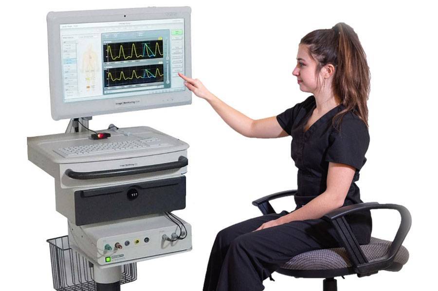
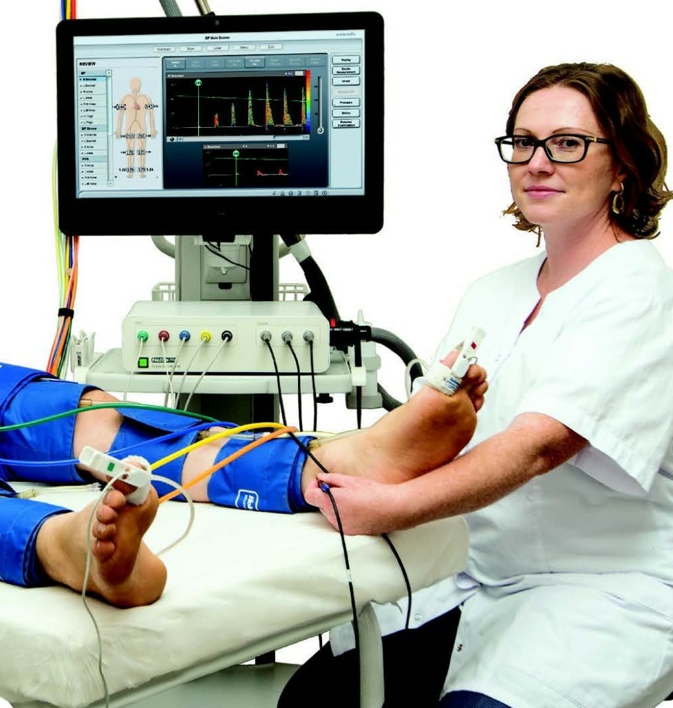
The FALCON PRO is the world’s most advanced high-end peripheral vascular diagnostic system. It is the ideal system for any vascular/angiology laboratory, and as an upgrade to existing old equipment.
Dolphin IQ and Dolphin 4D TCD Testing Systems
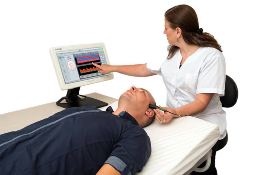
The next generation Transcranial Doppler (TCD) standalone system that will exceed everything you know and expect from a TCD system. The DOLPHIN supports numerous standard and new unique features for the benefit of the medical staff and patients alike!
Types of test conducted with a non-invasive diagnostic testing system
There are three main modalities used with a noninvasive diagnostic testing system.
Segmental blood pressures:
Segmental blood pressures is a physiologic test, which is performed using a physiologic machine in order to help in localizing arterial obstruction to flow along the limb, as well as the physiological severity of the obstruction. This is in contrast to ultrasound imaging of the lower limb, which is focused on detecting and quantifying the anatomical severity of the arterial obstruction to flow.
PVR or pulse volume recording:
Pulse Volume Recording is an air plethysmography waveform analysis test used to detect the segmental volume changes in the limb resulting from the flowing blood as a function of the cardiac cycle. This test is usually performed with a PVR machine.
PPG or photoplethysmography:
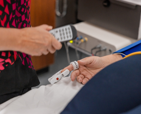
The PPG sensors transmit infra-red waveforms to the skin and detect the signals that are reflected back to the sensor from the skin. The skin partially absorbs the transmitted signals, and the reflected signals are a function of this light absorption, which in turn is a function of the local blood perfusion. Therefore, the PPG waveforms reflect local and relatively shallow skin variations in blood flow.
The Photo-plethysmograph detected local blood volume changes generate a pulsatile waveform, which is very similar to the Pulse Volume Recording (PVR) waveforms. However, PPG is based on optical technology, while PVR technology is based on a pressure-sensor measurement.
Other testing modalities you can test with the Falcon Pro and Falcon Quad:
- Doppler measurements
- Penile function test
- Reactive hyperemia
- Air plethysmography
- Raynads syndrome
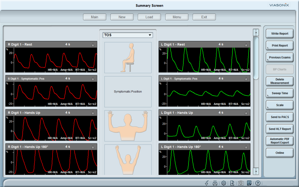
- Thoracic outlet syndrome (TOS)
- Palmar arch test
- AV Fistula
- Toe brachial Index (TBI)
- Ankle Brachial index (ABI)
How to perform lower arterial diagnostic measurements?
- A vascular test is carried out by specially qualified technologists, which is then reported to a vascular medicine doctor who interprets the results.
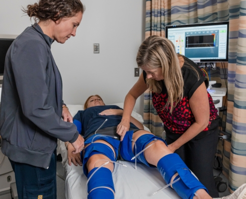
- During the test, the patient will be lying on a cushioned examination table.
- Depending on the test that has been ordered, he or she might spend some of the test walking on a treadmill.
- Depending on the body part being evaluated, multiple blood pressure cuffs are applied to various locations on the arms and legs.
- On the skin over the area that will be inspected, a little quantity of water-soluble gel is applied. Neither the skin nor clothing are harmed by this gel.
- A small instrument known as a transducer or doppler is placed on the skin’s surface and held there until the blood flow data has been recorded while the blood pressure cuffs are inflated.
- The tissues in the area being studied are employed by doppler to transfer sound waves.
- The interpreting doctor can determine the speed of the blood cells by using the sound waves reflecting them as they move inside the blood arteries.
Pulse Volume Recording Expected Results
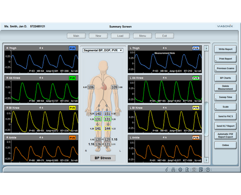
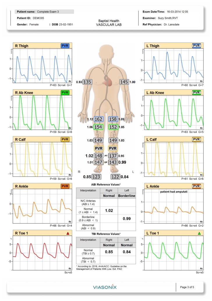
Pulse Volume analysis for diagnosis waveform recording is primarily qualitative in nature. The qualitative shape of the PVR waveform, notably the systolic rising curve, the maximum amplitude shape, the dicrotic notch during the descending region, and the waveform’s diastolic section, are typical of the doctor’s interest to plan further treatment. But it also offers a variety of quantitative parameters, such as waveform amplitude, rising time, or heart rate.
Pulse Volume Recording Normal Values
The components of a “normal” PVR waveform are a quick systolic upstroke, a moderately sharp peak, a downstroke with a noticeable dicrotic notch, and distinct diastole.
Pulse Volume Recording Interpretation
Your doctor or other healthcare professional compares the blood pressure in your both legs and arms. You can have a vascular disease if the blood pressure in your legs is lower than in your arms. To pinpoint the overall position of blockages or restricted arteries, your doctor may also compare the pulse in other areas of your leg.
FAQs
How do you record the volume of a pulse?
Pulse Volume Recording is an air plethysmography waveform analysis test used to detect the segmental volume changes in the limb resulting from the flowing blood as a function of the cardiac cycle.
What is a pulse volume recording test?
Pulse Volume Recording (PVR) is a pneumo-plethysmography test that is widely used for non-invasive diagnosis of lower extremity peripheral arterial disease (PAD).


